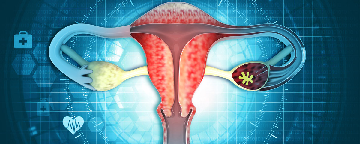
Endometriosis is derived from the word “endometrium” the tissue that lines the uterus. Women with endometriosis have endometrial tissue outside of the uterus, primarily the pelvic cavity, ovaries, fallopian tubes, and bowel (rectovaginal septum). Affecting 6-10% of women of reproductive age, endometriosis is characterized by dysmenorrhea (painful periods), dyspareunia (painful intercourse), chronic pelvic pain, irregular uterine bleeding and/or infertility. The prevalence of this condition in young women experiencing pain, infertility, or both is as high as 35-50%
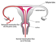
According to the most convincing model, the “retrograde menstruation hypothesis”, endometrial tissue fragments reach the pelvic cavity via trans fallopian tube retrograde (backward) flow, implant onto the peritoneum. Medical therapy is symptomatic, not cytoreductive (removal the abnormal tissue); and cytoreductive surgery is associated with high recurrence rates. Although lesion eradication is considered a fertility-enhancing procedure, the benefit on reproductive performance is moderate. Assisted reproductive technologies (e.g. IVF) constitute a valid alternative. Endometriosis is also associated with a 50% increase in the risk of epithelial ovarian cancer.
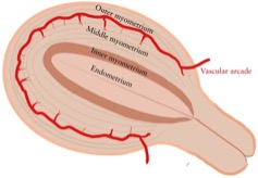
The inner myometrium is responsible for peristaltic-like, wavelike movements in the non-pregnant uterus. Hyperperistalsis and dysperistalsis (abnormal) of the uterus underlie many important disorders such as dysmenorrhea (painful menses) infertility, implantation failure and preterm birth.
The main function of the inner myometrium appears to be the modulation of uterine peristalsis, an area that increasingly is believed to play a significant role in fertility. Inner myometrial contractions vary in orientation, amplitude, and frequency throughout the menstrual cycle, under the influence of the sex hormones, estradiol and progesterone. Studies have shown that, in the follicular phase (early phase) of the cycle when the ovaries produce mature follicles which contain eggs, contractions are predominantly cervico-fundal (from the bottom of the uterus the cervix toward the top, specifically the fallopian tubes), and their amplitude and frequency increase significantly as ovulation approaches. There is evidence that this facilitates sperm ascension towards the distal (far) end of the fallopian tubes, where fertilization takes place. After ovulation, the uterus undergoes, under the influence of progesterone, progressive relaxation that culminates during the mid-luteal phase (second half of the menstrual cycle) which may assist proper embryo positioning in the midsection of the uterine cavity and, thus, facilitate embryo implantation while also improving the supply of oxygen and nutrients.
Women suffering from infertility in association with mostly mild endometriosis were subjected to vaginal sonography of uterine peristalsis during the menstrual period, the early, mid- and late follicular phases, and the mid-luteal phase of the menstrual cycle. The data obtained were compared with those of healthy controls. Women with endometriosis displayed a marked uterine hyperperistalsis that differed significantly from the peristalsis of the women without endometriosis (controls) during the early and mid- follicular and mid-luteal phases. During the late follicular phase of the cycle, uterine peristalsis in women with endometriosis became dysperistaltic (abnormal), arrhythmic and convulsive in character, while in controls, peristalsis continued to show long and regular cervico-fundal (normal) contractions.
This uterine dysperistalsis even in mild endometriosis, underlies many important disorders such as dysmenorrhea (pain), retrograde menses, and infertility due to interference with normal sperm transport.