Preterm birth, defined as delivery before 37 completed weeks of pregnancy, affects roughly one in ten pregnancies worldwide and remains the leading cause of neonatal mortality. Beyond the immediate risks to newborn survival, babies born too early face higher rates of long-term health challenges, including respiratory disease, neurodevelopmental delays, and chronic conditions that can persist into adulthood. Current preventive strategies, such as progesterone supplementation and cervical cerclage, are effective in carefully selected women, particularly those with a shortened cervix or a history of spontaneous preterm birth. Yet a major barrier persists: clinicians still struggle to accurately identify which women are truly at risk early enough for these interventions to have the greatest benefit.
For decades, transvaginal ultrasound measurement of cervical length has been the cornerstone of preterm birth risk assessment. A short cervix is one of the strongest known predictors, especially in women with symptoms of preterm labor or prior obstetric risk factors. However, large systematic reviews have shown that simply measuring cervical length has limited impact on reducing preterm birth rates at the population level. This gap has fueled interest in more advanced tools that can capture the complexity of pregnancy biology and better distinguish between women who will deliver early and those who will not.
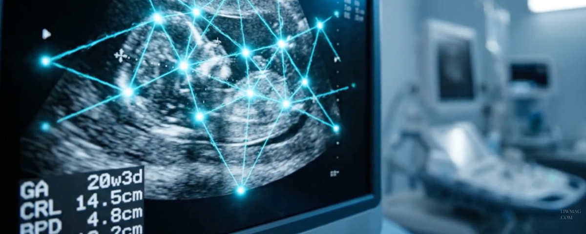
Recent research suggests that artificial intelligence (AI) may offer a meaningful step forward. By analyzing cervical ultrasound images using deep learning, a form of machine learning capable of recognizing complex patterns in raw image data, researchers have developed models that outperform cervical length alone in predicting spontaneous preterm birth. Unlike traditional approaches, AI does not rely solely on a single measurement. Instead, it evaluates subtle anatomical features, textures, shapes, and spatial relationships within the cervix and lower uterus that are difficult for the human eye to quantify consistently.
In a large multicenter study drawing on cervical ultrasound images collected over a decade from 19 hospitals; researchers trained and validated an AI model using thousands of real-world scans taken between 19 and 32 weeks of gestation. This dataset reflected everyday clinical practice, including variation in image quality, ultrasound machines, and operator experience. Importantly, the study focused specifically on spontaneous preterm birth, excluding medically indicated early deliveries, to ensure clinical relevance.
The AI model demonstrated significantly higher accuracy than cervical length measurement alone. It was better at identifying women who would deliver before 37 weeks, with particularly strong performance in predicting births before 34 weeks and, notably, late preterm births between 34 and 37 weeks; a group that accounts for the majority of preterm deliveries but is often missed by existing screening methods. At equivalent specificity, the AI approach detected substantially more at-risk pregnancies, meaning fewer women who would go on to deliver early were overlooked.
One of the most striking findings was that the AI model remained effective even when the cervix appeared “normal” in length. By using visualization techniques known as saliency heat maps, researchers found that the model often focused on regions of the lower uterine segment rather than the cervical canal itself. These areas are thought to reflect biomechanical and structural changes related to preterm birth risk, such as alterations in uterocervical angles and tissue properties; features clinicians have long suspected to be relevant but have struggled to integrate reliably into routine assessment.
The AI model also performed consistently across a wide range of maternal characteristics, including age, body mass index, parity, and prior obstetric history. Notably, it showed particular strength in women with higher BMI and in older pregnant women, groups for whom cervical length alone may be less predictive. Unlike cervical length, whose predictive value varies with gestational age, the AI model maintained robust accuracy even earlier in pregnancy. This is clinically important, as preventive treatments like progesterone or cerclage are most effective when initiated before 24 weeks.
From a practical perspective, the appeal of this approach lies in its seamless integration into existing care. The AI model analyzes standard transvaginal ultrasound images without requiring new scanning protocols or additional training for clinicians. By providing not only a risk estimate but also visual explanations of which anatomical features influenced the prediction, it has the potential to enhance clinical decision-making, improve patient counseling, and support more personalized care.
While these findings are promising, important limitations remain. The study was retrospective, and detailed information on symptoms, treatments, and some prior risk factors was not available for all participants. Prospective clinical trials are still needed to confirm whether improved prediction translates into better outcomes for mothers and babies. Nevertheless, this research represents a compelling proof of concept: AI can move preterm birth screening beyond a single measurement toward a more nuanced, individualized assessment of risk.
As reproductive and maternal health increasingly embrace precision medicine, AI-driven ultrasound analysis may become a powerful tool for identifying women who need closer monitoring or early intervention and just as importantly, for reassuring those who do not. For women navigating pregnancy or advising others, this work highlights how advances in data science and medicine are converging to address one of obstetrics’ most persistent challenges, with the potential to improve outcomes across generations.
Sources