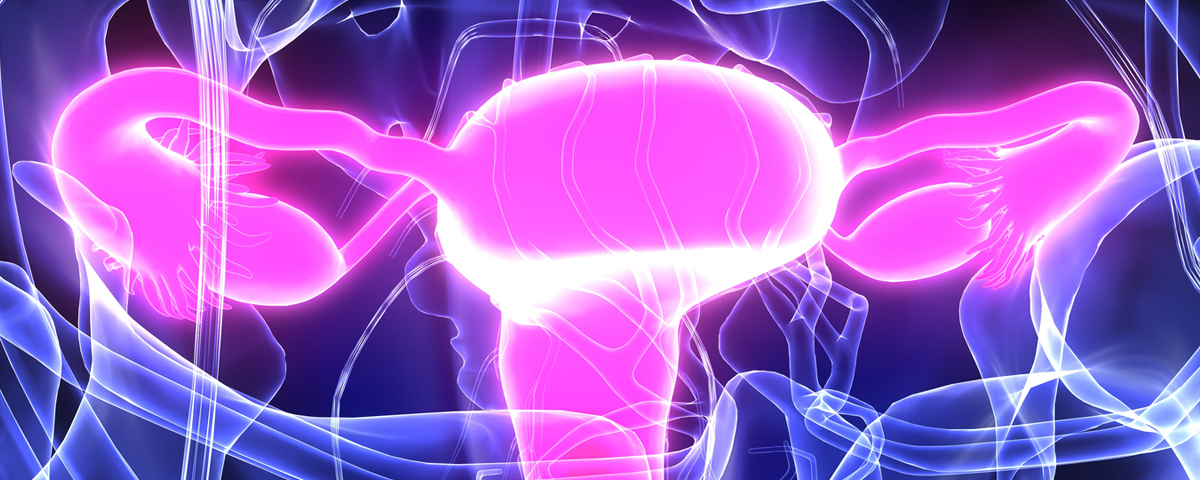
Reproductive aging in women is primarily due to the gradual reduction in the number of eggs (oocytes) over time. Oocytes cannot be reproduced, and while some develop and are released during ovulation, many others diminish as they become non-viable follicles and eventually disappear. This natural decline continues until menopause, at which point fertility ends entirely. Before menopause, fertility declines mainly due to the increased presence of oocytes with abnormal chromosomes (aneuploidy), which affects their viability.
With advancements in assisted reproductive technology (ART), how many oocytes can be collected through ovarian stimulation has become a focal point of discussion. While it’s true that the number of retrievable oocytes generally decreases with age, there’s a notable variation even among women of the same age. This is where the concept of “ovarian reserve” becomes important; it refers to the number of oocytes remaining in the ovaries. Currently, there is no direct way to measure this reserve, but certain serum hormone levels, such as follicle-stimulating hormone (FSH) and estradiol, are used as indirect indicators. However, this method has limitations as changes in these hormone levels often occur late in the process, just before ovarian function ceases.
The Anti-Müllerian Hormone (AMH) has emerged as a more reliable indicator in this context. Known for its role in fetal development, AMH is produced by the granulosa cells in ovarian follicles, and its levels can be measured in the blood. Although AMH doesn’t directly reflect the exact number of follicles in the ovary, it correlates well with the pool of growing follicles and, by extension, the remaining primordial follicles. Improvements in measuring AMH have enhanced its use as a test for ovarian reserve. Notably, AMH levels do not fluctuate significantly during the menstrual cycle, making it a stable and sensitive indicator of oocyte number decline, hence its growing clinical application over basal FSH levels.
AMH measurements now serve several clinical purposes, such as assessing fertility in the general population, predicting success in fertility treatments, diagnosing conditions like premature ovarian insufficiency (POI) and diminished ovarian reserve (DOR), and managing Polycystic Ovary Syndrome (PCOS). AMH levels are also useful for evaluation before and after ovarian surgery and during fertility preservation therapies.
Despite their value, AMH levels indicate the quantity, not the quality, of eggs and therefore don’t predict the likelihood of an egg leading to a successful pregnancy. For individual women, the number of remaining eggs serves as an important fertility indicator for a given time. When evaluating AMH levels’ utility, fertility outcomes are more telling than mere oocyte counts. Also, when AMH levels serve as diagnostic criteria, their relevance to specific diseases and diagnostic accuracy is crucial.
Summary of AMH and Women’s Reproductive Health
- General Population
- Fertility Assessment: AMH levels are only weak predictors of natural pregnancy. Low levels do not necessarily mean infertility. Their link to miscarriage risk is inconclusive.
- Menopause Prediction: AMH can suggest, but not precisely predict, when menopause will occur. Its best use is in combination with other measures, and more advanced testing might improve accuracy.
- Infertility Treatments
- Predicting Outcomes: AMH mainly helps predict success in women with low ovarian reserve. It doesn’t reliably forecast pregnancy for all women undergoing treatments like IUI or IVF.
- In Assisted Reproductive Technology ART: AMH can estimate ovarian response and likelihood of pregnancy, but prediction strength is moderate. Adjusting medication doses based on AMH can lower risks like ovarian hyperstimulation syndrome (OHSS) and optimize treatment.
- Premature Ovarian Insufficiency (POI) and Diminished Ovarian Reserve (DOR)
- Diagnosis and Prediction: AMH is highly effective for diagnosing POI early and predicting its onset, especially in women under 40. Low AMH levels indicate a higher risk, aiding in early intervention.
- Polycyclic Ovary Syndrome (PCOS)
- Diagnosis Aid: Elevated AMH levels are common in women with PCOS, making it a useful additional test for diagnosis. Variability in cutoff values depends on age and study methods.
- Surgical Impact on Ovarian Reserve
- Post-Surgery Monitoring: Measuring AMH before and after ovarian surgery helps evaluate how much ovarian reserve is lost, especially in procedures for ovarian tumors or endometriomas. A large decline in the ovarian reserve is more likely with bilateral surgery and certain surgical techniques.
- Fertility Preservation in Cancer Patients
- Post-Treatment Monitoring: AMH can indicate ovarian damage from chemotherapy or radiation, correlating with menstruation loss and ovarian reserve decline. However, its ability to predict future fertility after cancer treatment remains unclear, with some women conceiving despite low levels.
Conclusion:
AMH is a valuable tool for understanding ovarian reserve and guiding treatment in women facing fertility issues or disease-related ovarian damage. While useful in diagnosis, prediction, and treatment planning, its limitations mean it should be used alongside other assessments for best results. Ongoing research aims to enhance its predictive accuracy and clinical applications.