Urinary incontinence in women refers to the involuntary leakage of urine, affecting various aspects of their physical, psychological, and social well-being. It is a common condition, with over one in three adult women experiencing some form of urinary incontinence. While it’s more prevalent in older women, it can affect women of all ages and is often associated with life events unique to women, like pregnancy, childbirth, and menopause.
Types of Urinary Incontinence in Women:
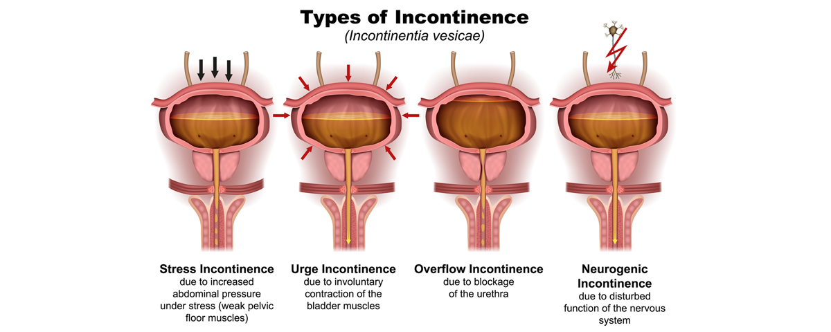
- Stress Urinary Incontinence (SUI): Urine leakage occurs during activities that increase abdominal pressure, such as coughing, sneezing, laughing, or exercising. This is often due to weakened pelvic floor muscles or damage to the urethra’s sphincter.
- Urge Urinary Incontinence (UUI): Characterized by a sudden, intense urge to urinate, often leading to leakage before reaching the toilet. It’s usually caused by overactive bladder muscles.
- Mixed Urinary Incontinence: A combination of both SUI and UUI symptoms.
- Overflow Incontinence: Occurs when the bladder doesn’t empty completely, leading to frequent small leaks. This is often due to an obstruction or blockage.
- Neurogenic Incontinence: a condition where the bladder doesn’t empty or store urine properly due to nerve damage or dysfunction.
The bladder, urethra and urinary sphincters work in concert to store urine at low pressure and to void voluntarily at socially convenient or appropriate times. The detrusor muscle and internal urethral sphincter are predominantly smooth muscle, whereas the external urethral sphincter and pelvic floor muscles are predominantly striated muscle. The bladder lumen is lined with epithelial cells (called urothelium) and the basement membrane (mucosal layer) which protect the underlying detrusor muscle from toxins contained in the urine and enable communication with neural cells that coordinate storage and voiding phases.
Voiding up to 7 times per day in the waking hours is considered normal, with a micturition volume of 250–300 ml per void (although the volume is typically higher with the first morning void). A healthy adult bladder has a limit of comfortable tolerance of approximately 500 ml and can accommodate this relatively large volume of urine with little, if any, increase in pressure, owing to the viscoelastic compliance of the bladder.
Two common, often overlapping, mechanisms for stress urinary incontinence have been described: urethral hypermobility resulting from loss of support of the bladder neck and urethra (such that they move during peaks of abdominal pressure), and weakness of the urinary sphincter itself. If the urinary sphincter mechanism is damaged, a specific subtype of stress urinary incontinence ensues, sometimes referred to as intrinsic sphincter deficiency. Weakness of the urinary sphincter can result from trauma, repeated uro -gynecological surgeries, neurological disease, ageing or diseases leading to systemic muscular atrophy.
Unlike the physical changes associated with stress urinary incontinence, urgency urinary incontinence involves physiological perturbations to bladder function. There are three main etiologies intrinsic to the bladder that can lead to urgency incontinence: detrusor overactivity, poor detrusor compliance and bladder hypersensitivity.
Detrusor Overactivity
Most current drugs for urgency incontinence block muscarinic receptors that mediate contraction within the detrusor muscle or stimulate βeta 3 receptors that promote relaxation. Many patients with urgency incontinence have measurable spontaneous uninhibited contractions of the detrusor muscle during bladder filling, called detrusor overactivity. These contractions can coincide with perceived urinary urgency, and urgency incontinence could result if the pressure generated overcomes the resistance of the urinary sphincter.
Poor Detrusor Compliance
In low compliance, the bladder fails to stretch, which causes increased pressure, discomfort during filling and a limited capacity. This pattern is typical after pelvic radiotherapy or can result from prolonged periods of catheterization.
Bladder Hypersensitivity
Afferent, sensory nerve impulses from the bladder to the Central Nervous System have recently been established as a key factor in overactive bladder syndrome and, accordingly, attention has focused on the sensory role of the urothelium in this condition. The urothelium is not merely a barrier but is a responsive structure that is able to detect thermal, mechanical and chemical stimuli.
With the recognition of the urothelium as a pivotal mediator of bladder function, much attention has been concentrated on the role of urothelial inflammation and infection in the etiology of overactive bladder syndrome. The balance of urinary microbiota is now believed to substantially alter bladder sensation and, perhaps, function. Historically, the bladder and urine were considered to be sterile, and urinary tract infection (UTI) in women was thought to result from ascending spread of uro-pathogenic organisms colonizing from the vagina. However, we now know there is a normal urinary microbiota dominated by lactobacillus.
The vaginal microbiota can be classified into five community-state types, of which four are dominated by different Lactobacillus species (L. iners, L. crispatus, L. gasseri and L. jensenii), whereas the fifth community is more diverse. Most urinary bacterial communities are also dominated by lactobacilli. Lactobacillus species especially in women is thought to be protective. The normal urinary microbiota help maintain bladder health and mucosal integrity. They also compete with or inhibit growth of uro-pathogens such as E. Coli and Klebsiella. They likely play a role in reducing the susceptibility to urinary tract infections UTI, overactive bladder and interstitial cystitis. Simply put women with UTIs often have decreased lactobacillus and increased uro-pathogens.
The management of urinary incontinence in adult women is an iterative process. For some affected women, urinary incontinence causes sufficient bother and intrusion to warrant consideration of treatment. The options range from lifestyle modification to more-invasive surgical interventions. Otherwise, healthy women might prioritize resolution of their urinary incontinence by actively engaging in pelvic floor rehabilitation, lifestyle changes (including fluid optimization), pharmacological treatment or surgery to resolve persistent symptoms.
Anticholinergic drugs (also known as antimuscarinics) are often used in primary care alongside behavioral and lifestyle modifications as part of first-line therapy. Anticholinergic drugs act directly on the detrusor muscle, which leads to reductions in urgency urinary incontinence, with concomitant improvements in urinary urgency, voiding frequency and, to a lesser extent, nocturia. Low-dose vaginal estrogen should be offered to post-menopausal women with urogenital atrophic changes, to promote improved blood supply.
Although conservative measures should be tried first, surgery to treat stress urinary incontinence is highly effective in reducing symptoms. Women with mixed incontinence are candidates for surgery but are likely to need adjunctive treatment for their urgency incontinence.