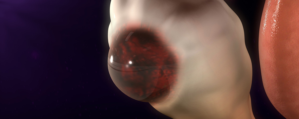
Endometriomas are cystic ovarian masses filled with old blood and tissue, stemming from endometriosis, a condition where tissue from the lining of the uterus, the endometrium grows outside the uterus. Endometriomas are often referred to as “chocolate cysts” due to the dark, reddish-brown fluid they contain. Endometriomas are a common finding in women with endometriosis and can indicate a more advanced stage of the disease. About 10% of young women will develop endometriosis, and about 17% to 44% of these women will also develop ovarian endometriomas and 28% of these women will have bilateral endometriomas.
Endometriomas are thought to develop from the invasion of endometrial tissue into the ovaries, or from the bleeding of the surface endometriosis into the ovarian cortex. While most commonly found in the ovaries, endometriomas can also occur in other locations, including the pelvic cavity, bowel, and bladder. The exact cause of endometriomas is not fully understood, but they are believed to be related to retrograde menstruation associated with endometriosis, where menstrual blood flows backward through the fallopian tubes and into the pelvic cavity.
The natural history of endometriomas is the same as endometriosis since endometriomas are a subset of this more extensive disease where ectopic endometrial tissue grows outside of the uterus. Ectopic tissue is tissue that grows outside of its natural habitat the lining of the uterus. The pathway for this disease process occurs with the hormonal response of the ectopic endometrial tissue. This tissue responds to the cyclical hormonal changes of a woman’s menstrual cycle in the same way that the intrauterine endometrium does. It will become proliferative grow, thicken and slough off at menstruation just as it would if it were within the uterus. Unlike the uterus where blood exits through the cervix and vagina during menstruation ectopic endometrial implants in the pelvic cavity bleed into the surrounding tissue. The blood has no outlet and accumulates locally, causing inflammation, irritation of the peritoneal surfaces, and pain. The repeated cyclical bleeding can also lead to fibrosis, scarring and adhesion formation.
With these fluctuations comes varying concentrations of cytokines and prostaglandin molecules. Cytokines and prostaglandins are signaling molecules for creating an inflammatory response and thus generate inflammation in the endometriotic implantation. This inflammatory response then lays the foundation for the production of new vascularization and new fibrous tissue formation. This snowball effect then creates the adhesions and pain commonly associated endometriosis and endometrioma. These issues also lead to the main complications of this disease, such as infertility and chronic pelvic pain. Patients with endometriomas have a more severe disease state and typically experience this on a more significant scale than those with stage one or two endometriosis.
Endometriomas can often be visualized on imaging. However, they appear similar to other cystic lesions on imaging, and final pathology is only discovered through surgery, normally laparoscopy where the chocolate cysts become apparent.
Endometriomas carry a risk of developing, epithelial ovarian cancers. A meta-analysis of 13 studies and approximately 9000 women with epithelial ovarian cancers showed that a self-reported history of endometriosis was three times as high.
Treatment of endometriosis mainly consists of hormonal medications or surgical treatment. Milder forms of endometriosis are treatable with oral contraceptive pills, various forms of progesterone (oral pill, intrauterine device), gonadotropin-releasing hormone (GnRH) agonists (such as leuprolide), or androgens (such as danazol). However, once a patient’s endometriosis has become severe enough to have the presence of an endometrioma, surgical management is typically preferred.
Ovarian cystectomy is a surgical procedure to remove endometriomas. The surgery typically involves laparoscopic excision, where the cyst wall is carefully removed, aiming to preserve as much healthy ovarian tissue as possible. This technique, also known as the “stripping” technique, is considered the standard treatment for endometriomas, especially those that are symptomatic or larger than 3 cm.
Although cystectomy is one of the most used approaches for the treatment of ovarian endometrioma, this endoscopic management is still controversial especially for women who plan to become pregnant. Several studies have suggested that ovarian reserve is compromised after laparoscopic cystectomy for ovarian endometrioma management. Ovarian reserve is defined as the functional potential of the ovary which reflects the number and quality of the remaining egg containing follicles.
Recent studies demonstrate that post operative hormonal treatment such as dienogest or GnRH may help preserve ovarian reserve in patients undergoing cystectomy for endometrioma. For women with infertility related to endometriomas, assisted reproductive technologies such as in vitro fertilization (IVF) may provide a pathway to conception.