Endometriosis and PCOS are the Yin and Yang of reproductive medicine. Endometriosis is driven by relatively low levels of prenatal and postnatal testosterone. Testosterone affects the developing hypothalamic–pituitary–ovarian (HPO) axis, and at low levels, it can result in an altered trajectory of reproductive and physiological phenotypes that can mediate the symptoms of endometriosis. Polycystic ovary syndrome, by contrast, is known to be caused primarily by high prenatal and postnatal testosterone, and it demonstrates a set of phenotypes opposite to those found in endometriosis.
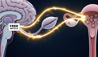
The hypothalamic-pituitary-ovarian (HPO) axis is a complex hormonal system that regulates female reproductive function. It begins with the hypothalamus, a brain region that secretes gonadotropin-releasing hormone (GnRH) in a pulsatile manner. GnRH stimulates the anterior pituitary gland to release two key hormones: follicle-stimulating hormone (FSH) and luteinizing hormone (LH). FSH primarily promotes the growth and maturation of ovarian follicles, which contain developing eggs, while also stimulating the production of estrogen by the granulosa cells within these follicles. LH plays a crucial role in triggering ovulation—the release of a mature egg—and supports the transformation of the remaining follicle into the corpus luteum, which secretes progesterone and some estrogen to prepare the endometrium for potential pregnancy. Estrogen exerts feedback regulation on the hypothalamus and pituitary, modulating the secretion of GnRH, FSH, and LH to maintain cyclical harmony. After ovulation, the corpus luteum produces hormones that sustain the early stages of pregnancy if fertilization occurs, or lead to menstruation if conception does not happen. This finely tuned feedback loop ensures the regulation of the menstrual cycle, fertility, and hormone balance throughout a woman’s reproductive years.
Early effects of prenatal testosterone, are important because they determine how and why the female HPO axis may become subject to early dysregulation that increases risks for endometriosis and other reproductive disorders.
Testosterone is present and active prenatally among females (at lower levels than among males), being produced from fetal and maternal adrenal glands, and maternal ovaries and maternal fat tissue, with maternal sources reaching the fetus via the placenta.
Among both females and males, prenatal testosterone levels have been linked with two main anatomical metrics. Anogenital distance (AGD), usually measured from the scrotal base to the anus in males, or from the distal end of the vagina to the anus in females, is much longer on average in males than females. Longer AGD is strongly associated with higher prenatal testosterone in both sexes, with considerable variation within each sex. A second metric of prenatal testosterone, the ratio of the 2nd to the 4th finger length (2D4D digit ratio) is lower among males than females, and lower digit ratio indexes higher prenatal testosterone within each sex as well, though with considerable variability. Some females thus develop under conditions of lower, or higher, prenatal testosterone than others, and lower prenatal testosterone involves a shorter AGD, longer 2D4D and notable effects on development of the HPO axis.
Endometriosis, in contrast to PCOS, exhibits a diverse set of hormonal indicators, correlates and risk factors suggesting that it is ‘opposite’ to PCOS in its causes and symptoms Most notably, women with endometriosis exhibit, relative to controls: shorter AGDs, indicative of lower prenatal testosterone (2) lower LH relative to FSH, (3) higher Sex Hormone Bonding Globulin SHBG, (4) higher serum oxytocin, (5) lower ovarian and serum testosterone, (6) lower Anti-Mullerian Hormone, AMH, (7) lower Waist to Hip Ratio WHR and BMI, (8) lower β-endorphin and (9) a suite of other differences, all of which are opposite to the differences found between PCOS versus controls.
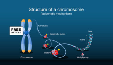
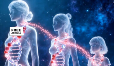
The epigenetics of endometriosis involves heritable changes in gene expression that do not alter the underlying DNA sequence but significantly influence the development and progression of the disorder. These modifications primarily include DNA methylation, histone modifications, and regulation by non-coding RNAs, such as microRNAs. In endometriosis, abnormal DNA methylation patterns have been observed in genes involved in hormone responsiveness, inflammation, cell proliferation, and apoptosis, leading to the aberrant survival and implantation of endometrial tissue outside the uterus. For example, hypermethylation of progesterone receptor genes can diminish tissue sensitivity to progesterone, contributing to progesterone resistance—a hallmark of endometriosis. Histone modifications may also alter chromatin structure and gene accessibility, further influencing the expression of genes involved in inflammation and angiogenesis. MicroRNAs regulate gene expression post-transcriptionally and have been found to be dysregulated in endometriotic lesions, affecting pathways related to immune response, cell migration, and invasion. These epigenetic alterations are often driven by environmental factors, such as toxins, stress, and hormonal imbalances, which can influence gene expression patterns across generations. Overall, epigenetic mechanisms contribute to the persistent inflammation, abnormal tissue growth, and heightened angiogenesis seen in endometriosis, offering potential targets for new diagnostic markers and therapies aimed at reversing epigenetic changes.
The epigenetics of polycystic ovary syndrome (PCOS) involves heritable changes in gene expression that do not alter the underlying DNA sequence but significantly influence the development and manifestation of the disorder. These epigenetic modifications include DNA methylation, histone modifications, and regulation by non-coding RNAs, such as microRNAs, which collectively affect genes involved in steroidogenesis, insulin signaling, inflammation, and ovarian function. In women with PCOS, abnormal DNA methylation patterns have been observed in key genes regulating androgen production, leading to hyperandrogenism, a hallmark feature of the disorder. For example, hypomethylation of genes like CYP17A1 can increase androgen synthesis, contributing to symptoms like hirsutism and acne. Additionally, altered histone modifications can change chromatin accessibility, affecting genes that control follicle development and insulin sensitivity, exacerbating reproductive and metabolic features of PCOS. Dysregulated microRNAs can further disturb gene networks involved in inflammation, insulin resistance, and ovarian follicle maturation. These epigenetic changes are influenced by environmental factors such as diet, stress, and toxins, which may trigger or worsen PCOS symptoms across generations. Understanding the epigenetic mechanisms underlying PCOS offers potential for developing targeted therapies aimed at reversing abnormal gene expression patterns, thereby improving reproductive and metabolic outcomes in affected women.