Polycystic Ovary Syndrome (PCOS) affects an estimated 8–13% of women of reproductive age. Up to 70% of cases are undiagnosed. There is slightly higher prevalence in Black and Middle-Eastern women than in the Chinese and White populations. Polycystic Ovary Syndrome is a metabolic and reproductive disorder characterized by high levels of androgens (male hormones), insulin resistance, and ovulatory dysfunction.
Symptoms and Diagnosis
PCOS changes manifest as hyperandrogenism (high levels of androgen) e.g. hirsutism (excessive hair growth), acne, or scalp hair loss, or a combination of these, and infrequent, irregular or absent menstrual periods. Long recognized as a reproductive disorder, polycystic ovary syndrome is now also established as a metabolic condition associated with long term health risks, including type 2 diabetes and cardiovascular disease. Adverse mental health outcomes and reduced quality of life have also been reported.
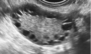
American Society for Reproductive Medicine (ASRM) concluded that PCOS should be defined (Rotterdam criteria) by the presence of two of the following three criteria: (1) oligo-ovulation (irregular menstrual cycles) or anovulation (ovaries do not release an egg); (2) clinical and/or biochemical signs of hyperandrogenism; (3) polycystic ovaries (PCO). PCO is defined using transvaginal ultrasound as the presence of 8 to 12 or more follicles in each ovary measuring 2-9 mm in diameter and/or increased ovarian volume (>10 mL).
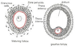
In normal women the wave theory holds that two or more cohorts of 4 to 14 small antral follicles ≥ 4 to 5 mm are recruited during the menstrual cycle and that one dominant follicle emerges in the final wave. The leading follicle can be distinguished from other members of the cohort by its size and the high mitotic index of the ovarian granulosa cells (cell division). Only the leading follicle has detectable levels of Follicle Stimulating Hormone (FSH) in the follicular fluid. The leading follicle also contains significant levels of estradiol. Selection does not always guarantee progression to ovulation but ovulation usually occurs.
Polycystic Ovary Syndrome PCOS is a misnomer, the disorder is not primarily a disorder of the ovaries but rather a neuroendocrine disorder. Neuroendocrine is defined as the interaction between the nervous system and the endocrine system.
Reproductive Physiology
The Hypothalamic-Pituitary-Ovarian (HPO) axis
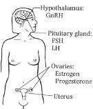
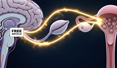
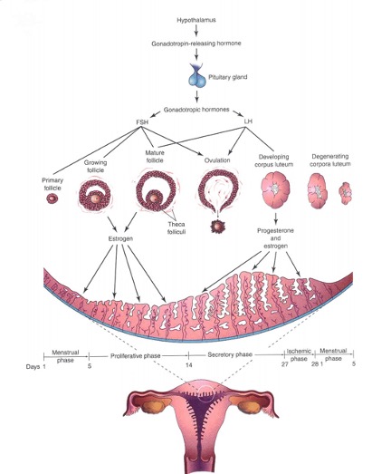
The female reproductive cycle (usually called menstrual cycle) is controlled by a neuroendocrine cascade involving the hypothalamus, pituitary gland, and ovaries (HPO axis). A neuronal clock located in the hypothalamus releases intermittent Gonadotropin Releasing Hormone (GnRH) that induces the release of the gonadotropins (hormones that stimulate the production of sex hormones estrogen and progesterone in the ovaries) specifically, Luteinizing Hormone (LH) and Follicle Stimulating Hormone (FSH) from the anterior pituitary. FSH and LH are responsible for the growth and maturation of the egg containing ovarian follicle via the timed production of estrogen and progesterone by the ovary (ovarian cycle). Estrogen and progesterone also promote cyclic changes in the endometrium, the lining of the uterus (menstrual cycle).
The neuronal clock (GnRH-containing neurons) located in the hypothalamus of the brain fires at regular hourly intervals, resulting in the intermittent release of gonadotropin-releasing hormone (GnRH). The GnRH is transported to the anterior part of the pituitary gland via the hypophyseal portal system
GnRH stimulates the cells of the anterior pituitary gland which in turn releases two hormones LH and FSH that will act on the ovaries: the gonadotropins (also called gonadotropic hormones) are Follicle stimulating hormone (FSH) and Luteinizing hormone (LH). Follicle stimulating hormone (FSH) stimulates the development of ovarian follicles and the production of estrogen by its follicular cells.
Luteinizing hormone (LH) is the trigger for ovulation, the release of an egg (oocyte). LH also stimulates the follicular cells and corpus luteum (a glandular structure that develops from the collapsed walls of the ovarian follicle shortly after ovulation) to produce progesterone and estradiol which are needed to maintain pregnancy should it occur.
& ENDOMETRIAL DEVELOPMENT
PCOS Pathogenesis
Alteration of the hypothalamic–pituitary–ovarian (HPO) axis is one major dysfunctions observed in PCOS; this modification is because of an increase in the pulse frequency release of the gonadotropin-releasing hormone (GnRH) from the hypothalamus, which, in turn, favors the synthesis and secretion of luteinizing hormone (LH) instead of follicle-stimulating hormone (FSH) by the pituitary gland. An elevation in serum levels of LH results in a high LH/FSH ratio (higher than2:1) that promotes androgen production in the ovarian theca cells.
Increased GnRH pulse generation in the hypothalamus increases LH pulse frequency, amplitude and as well as 24 hour men serum concentrations. Increased luteinizing hormone (LH) secretion together with enhanced ovarian theca cell responsiveness drives the production of excess androgen in PCOS.
Additionally, increased androgen production may inhibit sex steroid (estradiol/progesterone) negative feedback on hypothalamic Gonadotropin Releasing Hormone (GnRH) pulse generation resulting in the rapid LH pulse frequency seen in PCOS women. Increased androgens are also associated with android obesity, visceral fat deposition, and dyslipidemia (abnormal levels of lipids in the blood), all of which may contribute to insulin resistance and compensatory hyperinsulinemia. Hyperandrogenemia, obesity, and hyperinsulinemia and decrease Sex Hormone Binding Globulin (SHBG), increase free, bioactive androgens further driving the disorder.
Currently PCOS is treated symptomatically with oral contraceptives, antiandrogens, and insulin sensitizers. In women not attempting to conceive, combined contraceptive pills are first line treatments for menstrual irregularity and hyperandrogenism. The estrogen component increases sex hormone binding globulin, thus reducing androgenic free testosterone, and improving hyperandrogenism. Because this stimulatory effect on hepatic production of proteins also causes hypercoagulability, ethinylestradiol based contraceptive pills containing the lowest effective dose of oestrogen (e.g. 20-30 μg of ethinylestradiol) are recommended.
Currently available anti-androgen agents act by blocking androgen receptors (cyproterone acetate, spironolactone, and flutamide) or reducing production of androgens (finasteride and dutasteride).
Metformin modulates hepatic insulin sensitivity and glucose production. More recently, metformin has also been shown to mediate its anti-glycemic effects by actions on the gastrointestinal tract and the gut microbiome. Metformin is used to manage weight and metabolic outcomes in adult women with polycystic ovary syndrome with a body mass index ≥25. Metformin may also improve ovulation and live birth rates.
An improved understanding of the pathogenesis of PCOS will result in the development of new treatments and better patient outcomes. Recent studies have advanced our understanding of these pathophysiological processes. Neuroendocrine dysregulation leads to abnormal high frequency pulsatile secretion of gonadotrophin releasing hormone (GnRH), and hypothalamic kisspeptin, neurokinin B, and dynorphin A neurons (so-called KNDy neurons) are integral regulators of this process. Although increased production of ovarian and adrenal androgens contributes to hyperandrogenism, peripherally generated 11-oxygenated androgens are emerging as important predictors of metabolic risk. Together with advances in our understanding of adipocyte (fat storing cells) biology, insulin resistance, the gut microbiome, and insights from genome-wide association studies, these studies could improve our understanding of the pathogenesis of this disease. New treatments based on these observations are now in various stages of preclinical or clinical development.
One example of a new approach is inhibition of the neurokinin 3 receptor that is believed to cause a tempered inhibition of gonadotrophin releasing hormone (GnRH) pulsatility without excessive reduction because of the presence of compensatory pathways. A second approach takes advantage of the fact that when dynorphin binds to kappa opioid receptors, release of kisspeptin onto gonadotrophin releasing hormone neurons is inhibited, and therefore selective kappa receptor agonists with a central action might reduce gonadotrophin releasing hormone (GnRH) pulsatility. Additionally, kisspeptin receptor antagonists have also been suggested as therapeutic agents in PCOS based on their potential to normalize hypersecretion of luteinizing hormone (LH), restore folliculogenesis (the process by which a fertilizable egg is developed) and ovulation, and improve ovarian hyperandrogenism.
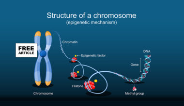
Lastly, epigenetic factors have garnered considerable attention in the pathogenesis of PCOS considering that changes in the content of DNA methylation, histone acetylation and noncoding RNAs have been reported in various tissues of women with this disease. Epigenetic changes are chemical modifications to DNA that regulate whether genes are turned on or off. DNA methylation is changed in the peripheral and umbilical cord blood, as well as in ovarian and adipose (fat) tissue of women with PCOS, suggesting the involvement of this epigenetic modification in the pathogenesis of the disease. Perhaps, these defects in DNA methylation promote the deregulation of genes involved in inflammation, hormone synthesis and signaling and glucose and lipid metabolism. Research on the role of DNA methylation in the pathogenesis of PCOS is just beginning, and several issues await investigation.
Sources