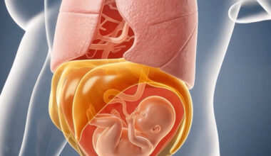

Endometriosis is classically defined as the presence of endometrial tissue both endometrial glands and stroma (connective tissue), in ectopic locations outside of the uterus, primarily the ovaries, fallopian tubes, pelvic peritoneum and rectovaginal septum. Affecting 6-10% of women of reproductive age, endometriosis is characterized by dysmenorrhea, chronic pelvic pain, irregular uterine bleeding and/or infertility and is occasionally accompanied by painful intercourse, bowel movements and/or urination. The prevalence of this condition in women experiencing pain or infertility, or both is as high as 35-50%.
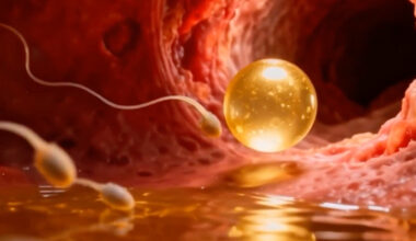
According to the most convincing model, the “retrograde menstruation hypothesis”, endometriosis occurs when menstrual fluid flows backward through the fallopian tubes into the pelvic cavity instead of leaving the body through the vagina. The viable endometrial fragments are driven through the fallopian tubes, possibly by a pressure gradient originating from dyssynergic (abnormal) uterine contractions. This backward flow carries viable endometrial cells, which can implant on the surfaces of pelvic organs, such as the ovaries, fallopian tubes, and peritoneum. Once implanted, these cells respond to hormonal signals and continue to grow and shed cyclically, similar to the normal endometrial lining within the uterus. This hypothesis is supported by the fact that retrograde menstruation is a relatively common physiological phenomenon; however, not all women with retrograde flow develop endometriosis, indicating that other factors contribute to the disease. These may include genetic predisposition, immune system dysfunction, and environmental factors that allow these cells to adhere, invade, and develop a blood supply. While retrograde menstruation alone does not account for all cases of endometriosis, it remains a foundational theory that helps explain how endometrial cells reach ectopic sites.
Endometrial tissue from disease-free women does not exhibit aromatase activity. Aromatase is a crucial enzyme responsible for converting androgens, such as testosterone and androstenedione, into estrogens, specifically estradiol, within tissues. In the pathogenesis of endometriosis, aromatase expression is abnormally elevated in the ectopic endometrial-like tissue outside the uterus. This increased activity leads to local overproduction of estradiol, which is a potent estrogen that stimulates the growth, survival, and proliferation of endometrial and endometriotic cells. Elevated estradiol levels in ectopic lesions create a self-sustaining cycle, promoting inflammation, angiogenesis (new blood vessel formation), and tissue invasion, all of which exacerbate the lesions’ persistence and severity. Unlike normal endometrial tissue, which is regulated by systemic hormonal signals, the excess local estradiol in endometriosis acts independently, fueling lesion growth even when systemic estrogen levels are low, such as during menopause or after hormonal suppressive therapy. This overproduction of estradiol also increases inflammatory mediators and pain-sensitizing substances, leading to the chronic pelvic pain characteristic of endometriosis. Therefore, aromatase’s heightened activity and the resulting excess estradiol are central to the development and progression of endometriotic lesions, making them important targets for therapeutic intervention to slow or halt disease progression.
Progesterone also plays a complex and crucial role in the pathophysiology of endometriosis. In a typical menstrual cycle, progesterone helps regulate the growth and shedding of the endometrial lining. However, in women with endometriosis, there is often a phenomenon known as “progesterone resistance,” where the endometrial tissue outside the uterus responds inadequately to progesterone. This impaired response is believed to result from altered progesterone receptor expression and signaling pathways within the ectopic endometrial tissue. As a result, the ability of progesterone to inhibit estrogen-driven proliferation and inflammation is compromised, leading to persistent growth and survival of these lesions. The reduced effectiveness of progesterone allows for an inflammatory milieu, contributing to pain and the formation of adhesions. Treatment often involves hormonal therapies that attempt to modulate this imbalance, such as progestins or oral contraceptives, which aim to enhance progesterone’s effects and suppress ovulation and reduce estrogen production. Understanding progesterone’s role and addressing the resistance mechanisms are key to developing more effective treatments for endometriosis, aiming to alleviate symptoms and improve fertility outcomes.
Endometriosis is an epigenetic disorder. Epigenetics is the heritable changes in gene expression that occur without alterations in the underlying DNA sequence. These changes influence how genes are turned on or off, effectively regulating cellular functions and development. Epigenetic modifications include mechanisms such as DNA methylation, histone modification, and the action of non-coding RNAs, which alter the structure and accessibility of chromatin—the complex of DNA and proteins in the nucleus. These modifications can be influenced by environmental factors like diet, stress, toxins, and lifestyle, and can be passed from one generation to another. For example, hypermethylation of progesterone receptor genes can diminish tissue sensitivity to progesterone, contributing to progesterone resistance—a hallmark of endometriosis. Histone modifications may also alter chromatin structure and gene accessibility, further influencing the expression of genes involved in inflammation and angiogenesis. MicroRNAs regulate gene expression post-transcriptionally and have been found to be dysregulated in endometriotic lesions, affecting pathways related to immune response, cell migration, and invasion. These epigenetic alterations are often driven by environmental factors, such as toxins, stress, and hormonal imbalances, which can influence gene expression patterns across generations. Overall, epigenetic mechanisms contribute to the persistent inflammation, abnormal tissue growth, and heightened angiogenesis seen in endometriosis, offering potential targets for new diagnostic markers and therapies aimed at reversing epigenetic changes
Standard diagnosis of endometriosis is carried out by direct visualization and histologic examination of lesions. Pain can be treated by surgically excising peritoneal implants, deep nodules and ovarian cysts, or inducing lesion suppression by abolishing ovulation and menstruation through hormonal manipulation with progestins, oral contraceptives and gonadotropin-releasing hormone agonists or antagonists. Medical therapy is symptomatic, not cytoreductive meaning all of the abnormal tissue is not removed. Surgery is associated with high recurrence rates. Although lesion eradication is considered a fertility-enhancing procedure, the benefit on reproductive performance is moderate. Assisted reproductive technologies such as IVF constitute a valid alternative.