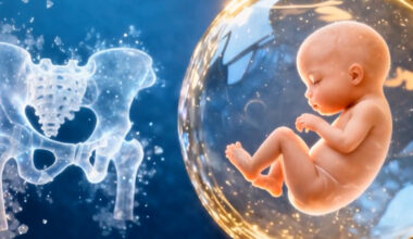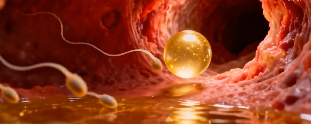
Endometriosis is a significant factor contributing to infertility in women, primarily due to its impact on the reproductive organs and functions. The presence of endometriotic tissue outside the uterus causes chronic inflammation, which can interfere with normal ovarian and tubal function, disrupting the process of ovulation, fertilization, and embryo implantation. The inflammatory environment surrounding endometriotic lesions releases cytokines and other mediators that can impair ovarian follicle development, reduce oocyte quality, and hinder ovum release. Additionally, scarring, adhesions, and pelvic distortions caused by endometriosis can block or limit the fallopian tubes, preventing eggs and sperm from meeting, or impairing embryo transfer into the uterus. The abnormal immune response associated with endometriosis can also create a hostile environment in the pelvis, further reducing fertility. Even in women who technically ovulate and have normal eggs, the synchronization of the hormonal signals necessary for successful conception and implantation can be disrupted by the inflammatory and fibrotic changes. As a result, endometriosis significantly decreases fertility rates and poses a challenge for conception, often requiring surgical intervention and assisted reproductive technologies.
Endometriosis can significantly impact ovulation, contributing to infertility through various physiological disruptions. The inflammatory environment created by endometriotic lesions results in the release of cytokines, prostaglandins, and other inflammatory mediators, which can interfere with the normal hormonal signaling required for ovulation. This inflammation can affect the ovaries directly, impairing follicular development and maturation. As a result, the follicles may not develop properly, leading to anovulation or irregular ovulation cycles. Additionally, the chronic inflammation associated with endometriosis often leads to the formation of ovarian cysts, known as endometriomas, which can further disrupt normal ovarian function by damaging healthy ovarian tissue and affecting hormone production. The presence of endometriosis can also alter the balance of reproductive hormones such as estrogen and progesterone, which are crucial for the timing and regulation of the ovulation process. Moreover, endometriotic adhesions can physically distort the ovarian structure, hindering the release of the egg from the follicle. This multi-faceted impact on ovulation contributes significantly to the fertility challenges faced by women with endometriosis.
Conception, when sperm and egg meet, occurs in the distal (far end) of the fallopian tube, specifically in the ampulla which is the widest section of the tube. Endometriosis can significantly impact the fallopian tubes, which are essential for natural conception. The ectopic endometrial tissue associated with endometriosis secretes inflammatory mediators, cytokines, and growth factors that promote fibrosis and tissue growth, leading to the formation of dense adhesions around the tubes. These adhesions can cause the fallopian tubes to become caught or fixed in abnormal positions, impairing their mobility and function. As a result, the normal process of capturing the ovum after ovulation and transporting it toward the uterus may be hindered, reducing the likelihood of fertilization. Moreover, endometriosis-related inflammation can damage the lining of the tubes, causing swelling, narrowing, or blockages—particularly in the ampullary or isthmic regions—further obstructing sperm from reaching the egg or preventing the fertilized embryo from passing through. In severe cases, the tubes may become completely obliterated or adherent to surrounding tissues, dramatically lowering the chances of natural conception. This tubal damage often necessitates surgical intervention to remove adhesions or, in many cases, the use of assisted reproductive technologies like in-vitro fertilization (IVF) to bypass tubal factors and increase the chances of pregnancy.
Endometriosis can significantly interfere with the process of embryo implantation in the endometrium, the lining of the uterus, which is a critical step for achieving pregnancy. The ectopic endometrial tissue associated with endometriosis creates an inflammatory environment within the pelvis, releasing cytokines, prostaglandins, and other inflammatory mediators that can alter the uterine environment. This inflammation can impair the endometrial receptivity by disrupting the normal hormonal signaling and gene expression patterns necessary for creating a receptive uterine lining. Moreover, endometrial tissue in women with endometriosis often exhibits abnormal expression of integrins, cytokines, and growth factors essential for embryo attachment, making it less capable of supporting implantation. The presence of fibrotic tissue and adhesions caused by endometriosis also physically distort the uterus and impair the embryo’s ability to properly implant into the endometrial lining. Additionally, the local immune response in women with endometriosis can be dysregulated, leading to increased destruction of the embryo or failure to establish a stable connection between the embryo and the endometrium. All these factors combined result in a hostile environment that hampers implantation, thereby contributing to infertility in women with endometriosis.
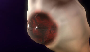
Abnormal Uterine contraction in Endometriosis is responsible for pain and infertility. The inner myometrium, the uterine muscle, is responsible for peristaltic-like, wavelike movements in the non-pregnant uterus. Hyperperistalsis and dysperistalsis (abnormal) of the uterus underlie many important disorders such as dysmenorrhea (painful menses) infertility, implantation failure and preterm birth.
The main function of the inner myometrium appears to be the modulation of uterine peristalsis, an area that increasingly is believed to play a significant role in fertility. Inner myometrial contractions vary in orientation, amplitude, and frequency throughout the menstrual cycle, under the influence of the sex hormones, estradiol and progesterone. Studies have shown that, in the follicular phase (early phase) of the cycle when the ovaries produce mature follicles which contain eggs, contractions are predominantly cervico-fundal (from the bottom of the uterus the cervix toward the top, specifically the fallopian tubes), and their amplitude and frequency increase significantly as ovulation approaches. There is evidence that this facilitates sperm ascension towards the distal (far) end of the fallopian tubes, where fertilization takes place. After ovulation, the uterus undergoes, under the influence of progesterone, progressive relaxation that culminates during the mid-luteal phase (second half of the menstrual cycle) which may assist proper embryo positioning in the midsection of the uterine cavity and, thus, facilitate embryo implantation while also improving the supply of oxygen and nutrients.
Abnormal uterine contractions even in mild endometriosis, can lead to Infertility.