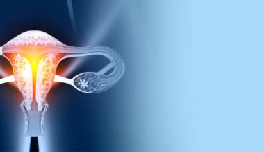
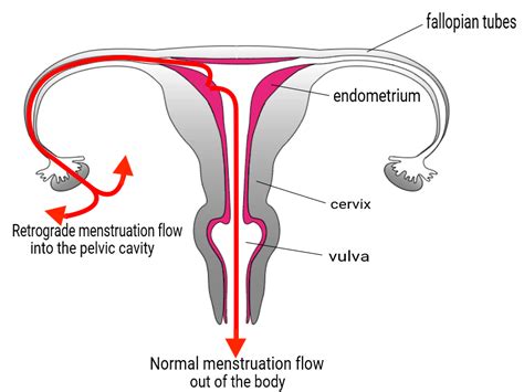
Endometriosis is a chronic, painful disease that affects around 10% of women of reproductive age worldwide. Retrograde menstruation is generally accepted as a cause of endometriosis, most women experience retrograde menstruation yet only 10% develop endometriosis, suggesting that other factors might contribute to its development.
(NORMAL VS RETROGRADE MENSES)
Fusobacterium infection of the endometrium (the lining of the uterus) may contribute to endometriosis. The reason why Fusobacterium preferentially infects the endometrium of some patients remains uncertain. Research has shown that Fusobacterium species originate in the oral cavity and are spread through the bloodstream to the uterine cavity especially, the endometrium. Such transmission mostly occurs during pregnancy, when blood flow to the placenta is generally increased. Because patients with endometriosis most often have not been pregnant, transmission through the vagina seems the more likely route in these women. The presence of Fusobacterium in the vaginal swab samples from patients with endometriosis was significantly greater than from patients without endometriosis, supporting the possibility of a vaginal transmission route.
Menstrual cycle-associated changes in mucin (natural protective proteins) and immune cell function, both barriers against bacterial infection, have been demonstrated in the female reproductive tract, which is associated with the individual’s susceptibility to infections. Fusobacterium appears to damage the mucin barrier and induce abnormal inflammation. These pathogenic roles of Fusobacterium may be due to its strong adhesion to vaginal tissue.
While the exact role of Fusobacterium in the pathogenesis of endometriosis is still being investigated, emerging evidence suggests that the bacteria may contribute to the development and progression of the disease through mechanisms involving inflammation, immune dysregulation, microbial dysbiosis (imbalance), tissue damage
, and hormonal influence. Further research is needed to fully understand the relationship between Fusobacterium and endometriosis and to explore potential therapeutic interventions.
The Mechanisms of Fusobacterium in Endometriosis Pathogenesis
1. Inflammation and Immune Response:Chronic Inflammation: Fusobacterium can induce chronic inflammation, which is a key factor in the development and progression of endometriosis. The bacteria can trigger the release of pro-inflammatory small proteins called cytokines, such as TNF-α, IL-1β, and IL-6, that play a crucial role in regulating immune response, inflammation, and cell growth and which can promote the growth and survival of endometrial-like tissue outside the uterus.
Immune Dysregulation: Fusobacterium may alter the local immune environment, leading to immune tolerance and allowing the ectopic endometrial (in an abnormal place) tissue to evade immune surveillance and destruction.
2. Microbial Dysbiosis (Imbalance):Vaginal and Endometrial Microbiome: Alterations in the vaginal and endometrial microbiome, including the presence of Fusobacterium, may contribute to the development of endometriosis. Dysbiosis can disrupt the normal microbial balance, leading to an environment that supports the growth of pathogenic bacteria and the development of endometrial lesions in various areas of the pelvis, such as the ovaries, fallopian tubes, bladder, and rectum.
3. Tissue Damage and Adhesion Formation:
Direct Tissue Damage: Fusobacterium can produce toxins and enzymes that directly damage the endometrial tissue, promoting the formation of adhesions and fibrosis, which are common features of endometriosis.Molecules: The bacteria may upregulate the expression of adhesion molecules on endometrial cells, facilitating their attachment to the ovaries, fallopian tubes, bladder, and rectum.
4. Hormonal Influence:
Estrogen Metabolism: Fusobacterium may influence estrogen metabolism, leading to increased local estrogen levels, which can promote the growth of endometriotic tissue. Estrogen is known to play a critical role in the pathogenesis of endometriosis.
With this greater understanding of the origination and development of endometriosis researchers are developing new therapeutic options.