Adenomyosis is a gynecological condition defined by the ectopic presence of endometrial glands and stroma, the tissue that normally lines the inside of the uterus, embedded within the myometrium, the muscular wall of the uterus itself. Unlike endometriosis, where similar tissue grows outside the uterus entirely, adenomyosis is contained within the uterine wall, causing it to become enlarged, thickened, and often globally distorted in architecture. The condition can be focal, presenting as a discrete adenomyoma (a nodular collection of ectopic endometrial tissue surrounded by hypertrophic smooth muscle), or diffuse, spreading throughout large portions of the myometrium. Clinically, it manifests as heavy menstrual bleeding (menorrhagia), severely painful periods (dysmenorrhea), chronic pelvic pain, and a characteristically enlarged, “boggy” uterus on examination. It is most commonly diagnosed in women in their 30s and 40s, though increasing use of high-resolution transvaginal ultrasound and MRI has revealed its presence in younger, even nulliparous women, challenging the old assumption that it was predominantly a disease of multiparous women.
The pathogenesis of adenomyosis remains incompletely understood, but several overlapping theories exist. The most widely accepted is the invagination or direct infiltration hypothesis, which proposes that the endometrial-myometrial junction (EMJ), a specialized zone of smooth muscle at the innermost layer of the myometrium, becomes disrupted, allowing endometrial cells to invade downward into the muscular wall. This disruption may result from mechanical trauma (such as uterine surgery, dilation and curettage, or cesarean sections), chronic inflammation, or intrinsic weakness of the junctional zone. A competing theory posits de novo metaplasia of mullerian remnants within the myometrium, while another proposes stem cell migration or tissue remodeling gone awry. Regardless of mechanism, once embedded, these ectopic glands and stroma respond to cyclic hormonal stimulation just as the normal endometrium does — proliferating under estrogen and cycling through secretory changes under progesterone — but without the ability to shed efficiently, leading to microhemorrhage, local inflammation, smooth muscle hyperplasia, and progressive fibrosis within the myometrium.
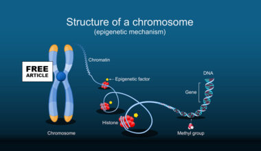
From an epigenetic standpoint, adenomyosis is increasingly understood as a disease of disordered gene regulation rather than straightforward genetic mutation. DNA methylation abnormalities are among the most well-characterized epigenetic changes: genes that should be silenced in normal myometrial tissue — including critical estrogen-signaling genes and inflammatory mediators — become hypomethylated and therefore aberrantly expressed in adenomyotic lesions. Conversely, genes that play protective or tumor-suppressive roles, such as progesterone receptor B (PR-B), undergo hypermethylation and are consequently silenced, contributing to the progesterone resistance that defines the disease phenotype. Histone modification patterns are similarly disrupted; altered acetylation and methylation of histones affect chromatin accessibility and shift gene expression profiles toward a pro-inflammatory, pro-proliferative state. Particularly striking is the epigenetic dysregulation of HOXA10 and HOXA11 — homeobox genes critical for uterine receptivity and endometrial function — which are abnormally expressed in adenomyotic tissue and impair normal uterine physiology. MicroRNAs (miRNAs) also play a significant regulatory role: several miRNAs are differentially expressed in adenomyotic tissue compared to normal endometrium, altering post-transcriptional regulation of genes involved in cell survival, invasion, and hormonal responsiveness. Crucially, many of these epigenetic marks are not fixed — they can be influenced by hormonal environments, inflammatory signals, and potentially by exposures that occur early in life or even in utero, opening a window into the developmental origins of the disease.
The in utero androgenic environment has emerged as a compelling area of investigation in understanding adenomyosis risk. The concept stems from the broader developmental origins of health and disease (DOHaD) framework, which holds that the hormonal milieu during fetal development can program tissue sensitivity and gene expression patterns that persist lifelong. Experimental animal models have been particularly instructive here: neonatal exposure to androgens or to diethylstilbestrol (DES, a synthetic estrogen) in rodents reliably induces adenomyosis-like uterine changes in adulthood, demonstrating that the uterus is exquisitely sensitive to hormonal programming during critical developmental windows. In humans, conditions associated with elevated fetal androgen exposure — such as polycystic ovarian syndrome (PCOS) in the mother, congenital adrenal hyperplasia, or exposure to endocrine-disrupting chemicals (EDCs) like bisphenol A (BPA) and phthalates — are associated with increased susceptibility to uterine pathology. Androgens influence the development of the Müllerian duct system and the differentiation of the junctional zone; aberrant androgen signaling during this period may alter the structural integrity of the EMJ or program epigenetic patterns that predispose to later invasion of endometrial cells into the myometrium. The androgen receptor (AR) is expressed in the uterus, and its downstream signaling intersects with estrogen pathways in ways that can either sensitize or desensitize tissue to subsequent hormonal stimulation, further linking fetal androgen levels to adult disease expression.
Adenomyosis is fundamentally an estrogen-dependent disease, and the enzyme aromatase (CYP19A1) is central to its self-sustaining hormonal pathology. Aromatase catalyzes the conversion of androgens — androstenedione and testosterone — into estrogens, primarily estradiol, and it is normally expressed at very low or undetectable levels in the healthy myometrium. In adenomyotic tissue, however, aromatase is aberrantly and robustly overexpressed, enabling the lesions themselves to generate their own local estrogen supply independent of the ovarian cycle. This intracrine and paracrine estrogen production creates a positive feedback loop: locally produced estradiol stimulates prostaglandin E2 (PGE2) synthesis via upregulation of cyclooxygenase-2 (COX-2); PGE2 in turn is one of the most potent known stimulators of aromatase expression, further amplifying local estrogen production, inflammation, and pain signaling. Meanwhile, progesterone — which normally counterbalances estrogen’s proliferative effects — is unable to exert its expected inhibitory influence because adenomyotic tissue is characterized by a profound reduction in progesterone receptor B relative to progesterone receptor A, a ratio shift that renders the tissue functionally resistant to progesterone. This dual pathology — excess local estrogen production driven by aromatase overexpression and simultaneous progesterone resistance — perpetuates a chronically estrogenic, pro-inflammatory, and pro-fibrotic environment within the uterine wall. It also provides the mechanistic rationale for treatments that suppress estrogen, including GnRH agonists and antagonists, combined hormonal contraceptives, progestins (though limited by the resistance), and the emerging use of aromatase inhibitors in refractory cases.
The impact of adenomyosis on fertility is multifactorial and increasingly recognized as clinically significant, particularly as more women delay childbearing. The mechanisms by which adenomyosis impairs reproduction are numerous and operate at virtually every stage of the reproductive process. At the level of the uterine environment, the architectural distortion of the myometrium and hypercontractility of the junctional zone disrupt the coordinated uterine peristaltic waves that are essential for sperm transport and embryo implantation. The normal endometrium — the lining of the uterus itself — is also functionally altered in adenomyosis, even though it is not the adenomyotic tissue per se: it demonstrates impaired expression of implantation markers such as integrins, pinopodes, and leukemia inhibitory factor (LIF), all of which are critical for embryonic attachment and invasion. The local inflammatory milieu — enriched in pro-inflammatory cytokines, reactive oxygen species, and prostaglandins — is hostile to both gametes and early embryos. Importantly, the aberrant epigenetic programming discussed earlier (including HOXA10 dysregulation) directly impairs endometrial receptivity during the implantation window. Women with adenomyosis who undergo in vitro fertilization (IVF) demonstrate significantly lower implantation rates, clinical pregnancy rates, and live birth rates compared to women without the condition, even when high-quality embryos are transferred — a finding that strongly implicates a uterine factor independent of ovarian reserve. Miscarriage rates are also elevated, likely reflecting both impaired implantation quality and the hostile uterine environment. While surgical treatment of adenomyosis (adenomyomectomy) can improve fertility outcomes in selected cases, it carries the risk of significant myometrial damage and uterine rupture in subsequent pregnancies, and medical suppression with GnRH agonists prior to IVF has shown promising but not yet definitive benefit in improving live birth rates. Adenomyosis thus represents one of the most complex intersections of endocrine pathology, epigenetic programming, and reproductive medicine, demanding both continued mechanistic research and individualized clinical management.